About Soft Tissue Shoulder Disorders
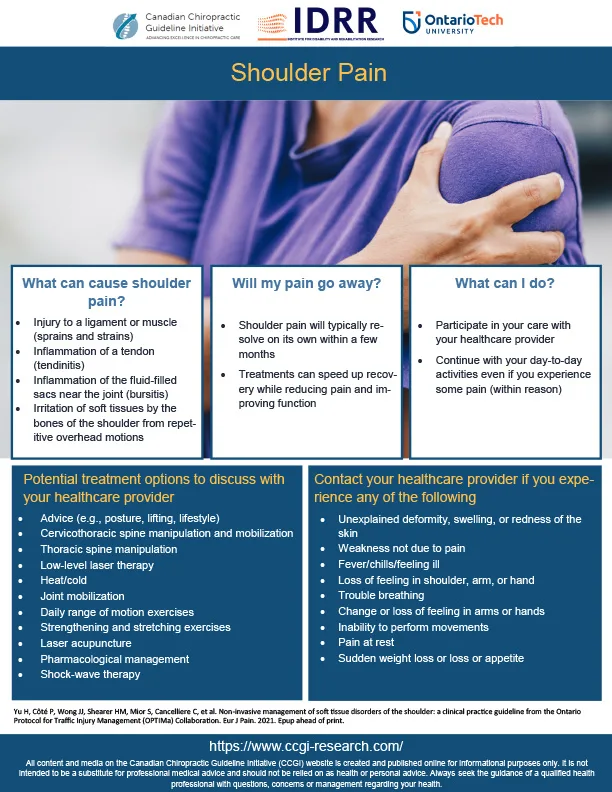
Shoulder pain is a common condition that can be acute or persistent, presenting as pain, discomfort and limited function in the shoulder area. Most cases respond well to conservative care, though some may result from serious underlying pathologies that require medical attention.
About CCG Care Pathways
Purpose
CCG care pathways provide structured, evidence-based guidance for clinicians delivering conservative, non-operative care for common musculoskeletal conditions. They outline key steps of the clinical encounter, support safe and appropriate decision-making, and assist with referral or co-management when indicated. Pathways are designed as practical, user-friendly tools that complement, not replace, clinical judgment.
Development
Pathways are developed using the best available evidence from high-quality clinical practice guidelines when they exist, and from systematic reviews and expert consensus when guideline evidence is limited or evolving. Content is reviewed periodically to reflect emerging research and current best practices. Input from clinicians, educators, and researchers helps ensure pathways remain relevant, aligned with real-world practice, and responsive to user needs.
Principles of Conservative Care
Musculoskeletal conditions are multifactorial and often influenced by physical, psychological, social, and environmental factors. As such, there is no one-size-fits-all approach to care. Effective management should be ethical, evidence-informed, transparent, flexible, and tailored to individual needs. Shared decision-making ensures care aligns with patient goals and values. Ongoing monitoring and outcome assessment support a person-centred approach and enable timely adjustments to care plans. Care may be delivered in-person, virtually, or through hybrid models, guided by patient preference, access, and clinical judgment.
Disclaimer
CCG care pathways are intended to support, not substitute for, professional clinical decision-making or the advice of a qualified healthcare provider. Recommendations are evidence-informed and presented in simplified, accessible language to support clinical understanding and application. Terms used throughout are not intended as formal diagnostic or billing terminology, nor are pathways prescriptive, authoritative, or regulatory.
Providers are expected to apply their clinical expertise and consult authoritative sources such as regulatory standards and policies, diagnostic classification systems (e.g., ICD-10-CA), scope-of-practice documents, continuing professional education resources, and peer-reviewed literature. Pathways may not apply to every clinical scenario and should always be interpreted in the context of individual patient needs.
Soft Tissue Shoulder Disorders Care Pathway
1. Record Keeping
Accurate, timely, and comprehensive documentation is an essential component of high-quality, evidence-based care. Clinical records must clearly reflect patient interactions, clinical reasoning, and progress over time, and should meet all jurisdictional regulatory standards.
Providers are encouraged to use a structured note format, such as the SOAP framework, to support consistency, clarity, and continuity of care.
Subjective: Document the patient’s reported symptoms, concerns, functional changes, contextual factors (e.g., psychosocial or environmental influences), and responses to prior care.
Objective: Record measurable or observable findings, including physical examination results, relevant diagnostic tests, functional assessments, and any clinically significant changes.
Assessment: Provide the clinical interpretation of findings, including diagnostic impressions or updates, identification of key risk factors or modifiers, and evaluation of the patient’s status or progression.
Plan: Outline the management strategy, including treatments delivered, modifications made, patient education and self-management recommendations, referrals, co-management decisions, and planned follow-up.
Documentation should be completed contemporaneously and maintained in accordance with regulatory requirements for privacy, security, and record retention. High-quality records support patient safety, facilitate interprofessional communication, enable shared decision-making, and promote continuity and accountability in care.
2. Informed Consent
- Definition: A process where the patient voluntarily agrees to proposed healthcare interventions after receiving adequate information on the nature, benefits, risks, and alternatives.
- Key Aspects:
- Prior to interaction: Obtain consent before any diagnostic testing or treatment. Ensure the patient understands the planned examinations, treatments, expected outcomes, and is given the opportunity to ask questions.
- Voluntarily and specific: Consent must be given willingly, without coercion, and pertain to the specific condition and proposed treatment. The patient should also understand that they can withdraw consent at any time.
- Transparent process: Consent must be obtained honestly, with a clear explanation of the condition and proposed interventions. Consent is not a one-time event, and involves ongoing discussions with the patient.
- Patient understanding and agreement:
- Diagnosis/prognosis: Explain findings clearly, using understandable language and visuals if needed.
- Treatment plan: Outline recommended treatments and how they align with patient goals. Discuss benefits, risks, and alternatives.
- Questions: Encourage questions and confirm understanding (e.g., “teach-back”).
- Documentation: Record the consent process, including information provided, patient questions, and explicit consent given.
3. Health History
- Apply cultural awareness and trauma-informed care principles.
- Sociodemographic information: Age, gender, sex, race/ethnicity.
- Main complaint: Location, onset, duration, radiation, frequency, intensity, character, aggravating/relieving factors, associated symptoms.
- Body systems review: Neurologic, cardiovascular, genitourinary, gastrointestinal, musculoskeletal, bone density, eyes/ears/nose/throat, respiratory, skin, mental health, reproductive.
- Health, lifestyle, and history: Past medical conditions, medications (including opioids, oral contraception, etc.), supplements, injuries, hospitalizations, surgeries, diet, exercise, sleep habits, smoking, alcohol/substance use, family support, caregiver responsibilities, work/school environment.
- Social determinants of health: Employment, childcare, education, nutrition, housing, domestic violence, child maltreatment, discrimination, social isolation.
- Previous treatments and responses: Document prior treatments, effectiveness, and any adverse effects.
- Beliefs and expectations: Assess patient understanding of their condition, treatment goals, and outcome expectations.
- Flag considerations: Identifyred, orange, and yellow flags for potential referrals.
Outcomes Assessments: Prioritize approaches that align with the patient’s specific goals and clinical presentation.
- Pain: Use pain scales (e.g., NRS) and diagrams.
- Function and Participation: Evaluate impact on daily activities (SPADI, DASH, QuickDASH, PSFS, WHODAS).
- Recovery: Use Self-rated recovery scales.
- Quality of Life: Assess using tools such as SF-12.
- Work/school Status: Monitor return to activities.
- Sleep quality: Assess using tools such as PSQI.
- Individual Goals: Set SMART goal setting (Specific, Measurable, Achievable, Relevant, Timely).
- Patient Feedback: Gather and integrate patient experience and satisfaction.
4. Red Flags : Differential Diagnosis Requiring Medical Referral
ACTION: Refer immediately to emergency care:
- Shoulder Infection: Immunosuppression, recent infection or surgery, TB (tuberculosis) history, unexplained fever/chills, IV drug use, poor living conditions.
- Traumatic Shoulder Fracture: Severe trauma.
- Acute Neurological Deficit (e.g., brachial plexus injury, acute cervical radiculopathy): Significant sensory/motor deficits in the upper extremity.
ACTION: Refer to appropriate medical provider:
- Shoulder Malignancy: Progressive pain, history of cancer, constitutional symptoms (fatigue, weight loss, night pain).
- Inflammatory Arthritides (e.g., polymyalgia rheumatica, rheumatoid arthritis, systemic lupus erythematosus): Morning stiffness >1 hour, systemic symptoms (fatigue, weight loss, fever), symmetrical joint pain, joint swelling and deformity, skin lesions.
- Referred Pain (from visceral conditions): Chest pain, shortness of breath, nausea (cardiac); shortness of breath, cough, pleuritic pain (pulmonary); epigastric pain, heartburn, post-meal pain (gastrointestinal); pain with deep breathing/coughing, hiccups (diaphragmatic).
5. Orange Flags: Symptoms of Psychiatric Disorders Requiring Referral
Clinicians should promptly address symptoms of potential mental health disorders to prevent harm through appropriate and timely referrals.
ACTION: Refer for immediate care (emergency department, medical/mental health provider):
- Suicidal ideation: Thoughts, plans, or statements about suicide or feelings of hopelessness.
- Severe, acute symptoms: Acute psychological distress, such as psychosis, severe panic.
- Ideation of harm: Intent or plans to self-harm, commit violence, or harm others.
ACTION: Refer to appropriate medical/mental health provider:
- Persistent, non-urgent symptoms: Symptoms affecting daily functioning (e.g., low mood, anxiety, sleep disturbances, social withdrawal, substance use).
ACTION: Co-management by non-medical/mental health providers:
- Triage: Ensure primary management by medical/psychiatric providers.
- Musculoskeletal (MSK) treatment: Manage MSK conditions related to or comorbid with psychological disorders.
- Screening tools: Selectively use tools to monitor symptoms and severity, guide care, and support escalation without implying a diagnosis. Tools include:
6. Yellow Flags: Psychosocial Factors that May Delay Recovery
Non-health barriers can delay recovery; early identification and intervention can enhance outcomes.
Factors:
- Individual: Worry, fear of movement, low recovery expectations, limited self-efficacy, reliance on passive treatments, activity avoidance.
- Social: Lack of family/social support, limited connections.
- Socioeconomic: Employment status, financial stress, litigation/compensation.
- Environmental/cultural: Social inequality, unsafe/unsupportive environments.
- Life events: Major transitions (e.g., divorce, job loss), chronic stressors (e.g., caregiving).
- Work/school: High stress, poor work-life balance, limited accommodations for injury/illness.
ACTION: Co-management by non-medical/mental health providers:
- Education & self-care: Provide resources for (e.g., stress management, coping strategies, graded activity).
- Monitor & coordinate: Regularly assess psychosocial challenges; refer to medical/mental health provider if persistent.
- Screening tools: Selectively use tools to monitor symptoms and severity, guide care, and support escalation (aligned with Orange Flag guidance), without implying a diagnosis. Tools include:
7. Physical Examination
- Observation: Evaluate abnormalities, asymmetries, posture, balance, gait, movements, facial expressions.
- Range of Motion (ROM): Assess active, passive, resisted shoulder ROM in flexion, extension, abduction, adduction, internal and external rotation).
- Motor strength: shoulder flexion, extension, abduction, adduction, internal rotation, external rotation, horizontal abduction, horizontal adduction.
- Palpation: Examine bone, joint, and muscle for tenderness, swelling, muscle tightness, or temperature changes.
- Neurological Examination:
- Motor strength testing: Assess for asymmetry or weakness in key muscle groups:
- Sensory testing: Assess for sensory deficits in dermatomal distributions:
- C4: Anterior shoulder
- C5: Lateral shoulder
- T1: Medial side of the forearm and upper arm
- T2: Medial aspect of the upper arm
- Reflex testing: Assess for asymmetry, diminished/absent reflexes:
- C5: Biceps brachii
- Upper motor neuron signs: Asses for increased muscle tone, hyperreflexia, pathological reflexes (e.g., Babinski sign, Clonus). May indicate central nervous system disorders (e.g., myelopathy, multiple sclerosis, stroke).
- Lower motor neuron signs: Assess muscle atrophy, fasciculations, reduced muscle tone, symmetrical loss of function. May indicate systemic neurological conditions (e.g., radiculopathy, peripheral neuropathy, ALS).
- Special/Orthopedic Tests: Perform as clinically indicated.
- Advanced Diagnostics: Radiography is generally not recommended without red flags or specific individual factors (e.g., contraindications to treatment).
8. Clinical Presentations for Soft Tissue Shoulder Disorders
Includes rotator cuff conditions (e.g., tendinopathies, partial tears), impingement syndrome, bursitis, calcific tendinitis, biceps tendon conditions, shoulder instability, AC joint conditions, labral tears and SLAP lesions, adhesive capsulitis, myofascial pain syndrome, osteoarthritis.
- Definition: Soft tissue shoulder disorders that are not due to serious underlying pathology requiring medical attention such as infection, tumor, or fracture, and are typically amenable to conservative care (e.g., education, manual therapy, exercise).
- Prevalence: Shoulder pain is a common musculoskeletal complaint in primary care, following back pain and knee pain.
- Pain: Localized shoulder area with or without referred pain.
- Signs/Symptoms: Sharp, dull, shooting, or aching pain; aggravated by specific movements; may refer into the arm.
- Exam: Pain reproduced by tests; typically, no neurological deficits.
Soft tissue shoulder disorders represent the most common causes of shoulder pain, presenting with similar mechanisms, clinical symptoms, and signs in a primary care setting.
Rotator Cuff Conditions (including tendinopathies and partial tears)
- Common in the general population, increasing with age.
- A dull ache in the lateral aspect of the shoulder, worsening with overhead activities and at night, weakness, limited range of motion, crepitus, night pain.
- Pain worsens with overhead movements, lifting, or reaching behind the back and improves with rest or activity modification.
- Exam findings include positive tests (e.g., drop arm, Jobe’s, infraspinatus, lift-off, belly press); no neurological deficits.
Impingement Syndrome
- Common condition that increases with age.
- Pain in the front or lateral aspect of the shoulder, particularly during overhead activities; shoulder muscle weakness; limited range of motion; night pain; crepitus.
- Pain worsens with overhead activities, reaching, or lifting and improves with rest or avoiding aggravating movements.
- Exam findings include positive tests (e.g., Neer’s, Hawkins-Kennedy, painful arc, Jobe’s, infraspinatus); no neurological deficits.
Calcific Tendinitis
- Affects approximately 3 to 8% of the general population, with higher prevalence in people aged 30 to 60 years.
- Sharp, intense pain in the shoulder, especially with movement; tenderness; limited range of motion; night pain; swelling.
- Pain worsens with shoulder movement, especially overhead activities, and improves with rest, but may persist at night.
- Exam findings include positive tests include inspection (swelling, redness), palpation (tenderness), limited range of motion, Neer’s, Hawkins-Kennedy, imaging (e.g., x-ray, ultrasound); no neurological deficits.
Bursitis
- Common, particularly in individuals with repetitive shoulder movements.
- Pain with movement, tenderness, swelling, limited range of motion, night pain.
- Pain worsens with overhead movements, direct pressure on the shoulder, or repetitive activity and improves with rest or reduced movement.
- Exam findings include positive tests include Neer’s, Hawkins-Kennedy, inspection (swelling, redness), palpation (tenderness), limited range of motion; no neurological deficits.
Biceps Tendon Conditions
- Common, particularly in athletes and individuals with repetitive shoulder use.
- Pain in the bicipital groove, tenderness, weakness, swelling, limited range of motion.
- Pain worsens with overhead activities, lifting, or resisted elbow flexion and improves with rest or activity modification.
- Exam findings include positive tests include Speed’s, Yergason’s, palpation (tenderness), limited range of motion; no neurological deficits.
Shoulder Instability
- More common in younger, active individuals and athletes.
- Pain, sensation of the shoulder “slipping out,” weakness, limited range of motion, instability.
- Pain worsens with overhead movements, sudden arm motions, or carrying heavy objects and improves with rest or shoulder stabilization.
- Exam findings include positive tests (e.g., Apprehension, Relocation, Sulcus Sign); no neurological deficits.
Acromioclavicular (AC) Joint Conditions
- Common, particularly in athletes and older individuals.
- Pain at the AC joint, tenderness, swelling, limited range of motion, pain with horizontal adduction.
- Pain worsens with cross-body movements, overhead activities, or direct pressure on the joint and improves with rest or avoidance of aggravating movements.
- Exam findings include positive tests include Cross-Body Adduction, palpation (tenderness), inspection (swelling); no neurological deficits.
Labral Tears and SLAP Lesions
- Common in athletes and individuals with repetitive overhead activities.
- Pain, clicking or catching sensation, weakness, limited range of motion.
- Pain worsens with overhead movements, lifting, or throwing and improves with rest or reduced shoulder activity.
- Exam findings include positive test (e.g., O’Brien, Crank, Apprehension test, limited range of motion); no neurological deficits.
Adhesive Capsulitis
- Common, particularly in people aged 40 to 60 years, with a higher prevalence in women.
- Gradual onset of shoulder stiffness and pain, limited range of motion in all directions, pain worsening at night.
- Pain worsens with movement and at night, particularly when lying on the affected shoulder, and may improve with gentle mobilization.
- Exam findings include limited active and passive range of motion, particularly in external rotation; pain and stiffness during movement; positive Apley scratch test.
Myofascial Pain Syndrome
- Common, particularly in individuals with repetitive strain or stress.
- Tender trigger points in the muscles, muscle stiffness, referred pain patterns, limited range of motion.
- Associated with taut muscle bands and trigger points causing localized and referred pain upon compression.
- Exams include palpation to identify trigger points, assessment of referred pain patterns, limited range of motion; no neurological deficits.
Osteoarthritis
- Exams include inspection (swelling), palpation (tenderness), limited range of motion, crepitus during movement, imaging (e.g., X-ray); no neurological deficits.
- Common, particularly in older individuals.
- Pain, stiffness, limited range of motion, crepitus, swelling.
- Pain worsens with activity and improves with rest; morning stiffness may be present.
- Exams include inspection (swelling), palpation (tenderness), limited range of motion, crepitus during movement, imaging (e.g., X-ray); no neurological deficits.
9. Treatment Considerations for Soft Tissue Shoulder Disorders
Approach to Treatment
The treatments outlined in this section reflect core domains of care consistently identified across high-quality clinical practice guidelines and established clinical practices. These include interventions shown to improve patient-important outcomes such as pain, function, and quality of life. Management plans should be tailored to the individual’s needs, goals, and preferences, taking into account clinical presentation, response to care, and contextual factors.
Not all domains need to be included in every care plan or at every stage of recovery. Clinicians are expected to apply professional judgment in selecting the most relevant components based on the clinical context.
This pathway is not prescriptive, nor does it list every possible intervention. Readers are encouraged to consult individual guidelines for specific treatment protocols, dosage, and condition-specific considerations.
While a range of other interventions may be in use, such as passive physical modalities, these have mixed or limited evidence of clinical benefit and are therefore not recommended for routine use. If applied, such therapies should be used as adjuncts to the core, evidence-based components of care, and not as standalone treatment.
- Education and Self-Management (Yu et al., 2021, Lowry et al., 2024)
- Education & reassurance: Emphasize soft-tissue shoulder disorders’ often self-limiting nature. Use tailored, evidence-based information in various formats (written, digital, visual) to empower individuals. Limited evidence suggests no single superior type of education for improving patient outcomes, but consistent reinforcement improves understanding and engagement.
- Self-care: Encourage regular exercise, nutrition, sleep hygiene, stress management, weight maintenance, and avoidance of smoking/substance abuse. Employ SMART goals and Brief Action Planning for sustained engagement.
- Daily activities: Promote continued movement and discourage prolonged rest to enhance recovery and prevent functional decline.
- Social & work engagement: Encourage participation using pacing strategies and workplace accommodations to support social functioning and productivity.
Interventions for Shoulder Pain (Any Duration):
- Exercise therapy (Yu et al., 2021, Lowry et al., 2024)
Develop individualized programs focused on improving strength, mobility, and aerobic fitness, tailored to patient needs and preferences. Exercise has demonstrated benefits in reducing pain, improving functional capacity, and enhancing quality of life. No single exercise type is shown to be superior, so programs should align with patient capabilities and goals. Monitor psychological responses to exercise; refer to medical/mental health providers if signs of distress or aversion arise. - Manual Therapy (Yu et al., 2021, Lowry et al., 2024)
Incorporate spinal manipulation techniques to reduce pain and improve function. Manual therapy should be integrated as part of a broader care plan to maximize effectiveness.- Cervicothoracic/thoracic spine manipulation: For shoulder pain associated with restricted movement or pain in the cervicothoracic spine.
- Low-level laser therapy (Yu et al., 2021, Lowry et al., 2024)
- Utilize low-level laser therapy to reduce pain and enhance tissue healing. Low-level laser therapy should be integrated as part of a broader care plan, including exercise and patient education, to maximize effectiveness.
- Multimodal care (Yu et al., 2021, Lowry et al., 2024)
Integrate interventions tailored to individual needs, particularly to support function, work, and community engagement through predominantly non-pharmacologic care. - Medications (Yu et al., 2021, Lowry et al., 2024)
- Short-term relief: Consult a medical provider. Options may include short-term use of analgesics, NSAIDs, or muscle relaxants. Long-term opioid use is discouraged due to dependency risk.
- Corticosteroid injections (Lowry et al., 2024)
- For severe pain or functional limitations; for conditions like rotator cuff tendinopathy, adhesive capsulitis, or bursitis. Use short-term, with caution regarding long-term risks.
Additional Considerations for Chronic Shoulder Pain (>3 months):
- Laser Acupuncture (Yu et al., 2021, Lowry et al., 2024)
- If conventional treatments have not provided adequate relief.
- Shockwave therapy (Yu et al., 2021, Lowry et al., 2024)
- For calcific tendinitis based on imaging findings.
- General Physician Care (Yu et al., 2021, Lowry et al., 2024)
- Provide information, advice, and pharmacological pain management when necessary.
10. Risk and Prognostic Factors for Soft Tissue Shoulder Disorders
- Common Risk Factors: (Baumgarten et al., 2010; Cook et al., 2015; Dean & Söderlund, 2015; Dunn et al., 2014; Passaretti et al., 2015)
- Risk factors for shoulder pain share many similarities with other musculoskeletal conditions. These include genetic predisposition, hormonal influences, lifestyle factors (e.g., smoking, alcohol consumption, sedentary lifestyle), comorbidities, low level of education, sleep disorders, psychosocial factors (e.g., depression, anxiety, poor coping skills, and somatization). Work-related factors (e.g., excessive or maladaptive mechanical load on the tissues, repetitive task in overhead activities)
- Prognosis: (Littlewood et al., 2013; Luime et al., 2004; Murphy & Carr, 2010)
- Most individuals with soft tissue shoulder disorders recover, though recurrences are common. (Luime et al., 2004)
- Common negative prognostic indicators include (Littlewood et al., 2013; Murphy & Carr, 2010): Longer duration of symptoms, higher severity and complex types of injuries (e.g., full-thickness rotator cuff tears, labral tears, shoulder dislocations, severe osteoarthritis), older age, lower activity level and physical fitness, poor initial response to treatment, mental health issues, poor recovery expectations, work-related factors, previous shoulder injuries.
11. Ongoing Follow-up
- Monitor progress: Reassess symptoms, functional status, and patient-reported outcomes at appropriate intervals. Confirm that care remains aligned with the patient’s goals, values, and expectations.
- Adjust treatment plan: Continuously realign the management plan based on evolving goals, treatment response, clinical findings, and professional judgment. Modify interventions, dosage, frequency, or focus as needed to support meaningful improvement.
- Support self-management: Reinforce the patient’s understanding of home strategies, activity recommendations, and behavioural approaches. Encourage adherence and address barriers that may affect progress.
- Recognize plateaus or change in status: Identify when the patient is improving, stable, or worsening. Reassess for contributing factors such as comorbidities, psychosocial influences, or new functional limitations.
- Referral and co-management: Consider referral or co-management with an appropriate provider when there is limited or no significant improvement within an expected timeframe (for example 6 to 8 weeks), when new or concerning findings emerge, or when additional expertise is required to support optimal care.
- Documentation: Record follow-up assessments, changes to the plan, patient feedback, reassessment of goals, and any referral or co-management decisions.
12. Criteria for Discharge
- Discharge criteria: Establish clear criteria for concluding active care. These may include achieving the patient’s initial goals, demonstrating meaningful improvement in symptoms or function, reaching a plateau in progress, or transitioning to self-management as the primary approach. Consider patient preferences, functional demands, and clinical judgment when determining readiness for discharge.
- Clinical reassessment: Prior to discharge, complete a focused reassessment to confirm stability of symptoms, functional status, and the patient’s confidence in managing their condition. Address any remaining concerns and ensure no new issues require further evaluation.
- Post-discharge planning: Discuss ongoing self-management strategies, including activity recommendations, home exercises, behavioural or lifestyle modifications, and symptom monitoring. Provide guidance on when to return for follow-up, when to seek additional care, and what indicators should prompt medical evaluation.
- Future care needs: Clarify options for episodic care, preventive visits, or re-engagement with the provider if symptoms recur or functional demands change. Encourage ongoing communication if new concerns arise.
- Documentation: Record the rationale for discharge, the patient’s status at the time of discharge, self-management recommendations provided, and the agreed-upon follow-up plan
Exercise Videos
We would like to acknowledge our shoulder exercise panel members for their contributions: Dr. Nicholas Moser, Dr. Kim Castle, Dr. Caroline Poulin and Dr. Nadia Richer for their work in the conceptualization of these exercises. In particular, we would like to thank Drs. Poulin and Richer for their work in the development, translation, and demonstration of these videos. We would also like to thank Dr. André Bussières for narrating these videos and Mr. Yannick Maltais for recording and editing the videos.

Introduction to CCGI shoulder exercise videos
Shoulder mobility

Scapular mobility

Pendulum

Tables Wipes

Broomstick Mobility

Trapezius relaxation exercise
Shoulder stretching

Towel assisted stretch

Sleeper stretch

Posterior shoulder stretch

Pectoral stretch
Isometric shoulder exercises

Isometric shoulder exercises
Shoulder strengthening

Eccentric full can

Serratus punch

Push up plus

Eccentric external rotation

External rotation

Internal rotation

Low row

Straight arm pull down

Front Raises
Advanced

Subscapularis pull

Standing three ways

Modified empty can

Field goal
Patient Handout
References
- Baumgarten KM, Gerlach D, Galatz LM, Teefey SA, Middleton WD, Ditsios K, et al. Cigarette smoking increases the risk for rotator cuff tears. Clinical Orthopaedics and Related Research. 2010;468:1534-41.
- Cook JL, Rio E, Lewis JS. Managing tendinopathies. In: Jull G, Moore A, Falla D, Lewis JS, McCarthy C, Sterling M, editors. Grieve’s Modern Musculoskeletal Physiotherapy. 4th ed. London: Elsevier; 2015.
- Dean E, Söderlund A. Lifestyle factors and musculoskeletal pain. In: Jull G, Moore A, Falla D, Lewis JS, McCarthy C, Sterling M, editors. Grieve’s Modern Musculoskeletal Physiotherapy. 4th ed. London: Elsevier; 2015.
- Dunn WR, Kuhn JE, Sanders R, An Q, Baumgarten KM, Bishop JY, et al. Symptoms of pain do not correlate with rotator cuff tear severity: a cross-sectional study of 393 patients with a symptomatic atraumatic full-thickness rotator cuff tear. The Journal of Bone and Joint Surgery. 2014;96:793-800.
- Littlewood C, May S, Walters S. Epidemiology of Rotator Cuff Tendinopathy: A Systematic Review. Shoulder & Elbow. 2013 Oct 1;5(4):256-65.
- Luime JJ, Koes BW, Hendriksen IJ, Burdorf A, Verhagen AP, Miedema HS, Verhaar JA. Prevalence and incidence of shoulder pain in the general population; a systematic review. Scandinavian journal of rheumatology. 2004 Mar 1;33(2):73-81.
- Lowry V, et al. A systematic review of clinical practice guidelines on the diagnosis and management of various shoulder disorders. Arch Phys Med Rehabil, 2024.
- Passaretti D, Candela V, Venditto T, Giannicola G, Gumina S. Association between alcohol consumption and rotator cuff tear. Acta Orthopaedica. 2015:1-4.
- Murphy RJ, Carr AJ. Shoulder Pain. BMJ clinical evidence. 2010;2010.
- Yu, H. et al. Noninvasive management of soft tissue disorders of the shoulder: A clinical practice guideline from the Ontario Protocol for Traffic Injury Management (OPTIMa) collaboration. European Journal of Pain, 2021.